Understanding Function Brain Regions: What Actually Happens Up There
By Sarah Mitchell, Neuroscience Correspondent Published: November 2025
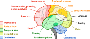
Function brain regions are basically the specialized neighborhoods of your brain where different types of processing happen. Think of it like a city – you wouldn’t go to the industrial district to watch a movie, right? Same deal with your brain. The occipital lobe handles vision, the temporal lobe deals with sound and memory, frontal areas manage decision-making and personality. But honestly, it’s way messier than most textbooks make it sound.
The Historical Discovery of Brain Localization
The whole concept of brain regions having specific jobs goes back to the 1860s when Paul Broca found that damage to a particular spot in the left frontal lobe (now called Broca’s area, obviously) screwed up people’s ability to speak. His patient, nicknamed “Tan” because that’s literally the only word the guy could say, had a lesion in what we now know as the left inferior frontal gyrus. Pretty wild that one case back in 1861 basically launched modern neuropsychology.
How Different Brain Regions Actually Work
Your brain isn’t just randomly organized. There are about 86 billion neurons up there (give or take a few billion – nobody’s actually counted them all individually), and they form these incredibly complex networks. The crazy part is that even though we talk about “function brain regions” like they’re separate departments, they’re constantly talking to each other through something like 100 trillion synaptic connections.
The Prefrontal Cortex and Executive Functions
The prefrontal cortex is probably the most talked-about region these days. It’s right behind your forehead and doesn’t fully develop until you’re around 25 years old, which explains a lot about why teenagers make questionable decisions. This region handles executive functions – planning, impulse control, working memory, that kind of stuff. Companies like Neurosky and Emotiv have been trying to measure prefrontal activity with consumer EEG headsets since around 2007, though the scientific community is pretty skeptical about how accurate those $99 devices really are.
The Hippocampus and Memory Formation
Then you’ve got the hippocampus, which looks like a seahorse (that’s literally what “hippocampus” means in Greek). This thing is crucial for forming new memories. The famous patient H.M. (Henry Molaison – they only revealed his real name after he died in 2008) had his hippocampus removed to treat severe epilepsy back in 1953. Afterwards he couldn’t form new long-term memories at all. He could remember stuff from before the surgery, could hold a conversation, but five minutes later he wouldn’t remember talking to you. Brenda Milner at McGill University studied him for decades.
Major Functional Areas You Should Know About
Motor and Sensory Cortex
Motor cortex sits in a strip across the top of your brain, right along the central sulcus. There’s this weird thing called the motor homunculus – if you mapped out which parts of the motor cortex control which body parts, you’d get this distorted little person with HUGE hands and lips and a tiny torso. That’s because fine motor control for things like your fingers and mouth needs way more neural real estate than, say, your elbow.
Wilder Penfield mapped this out in the 1950s at the Montreal Neurological Institute by literally poking people’s brains with electrodes during surgery (they were awake!) and seeing what moved. Can you imagine? “Hey doc, my thumb just twitched.” “Perfect, let me write that down.”
The sensory cortex is right next to the motor strip, just on the other side of that central sulcus. It receives touch, temperature, pain signals from your body. Again, you get this homunculus effect – your hands and face get disproportionate representation because they’re so sensitive.
Visual Processing in the Occipital Lobe
Visual processing happens primarily in the occipital lobe at the back of your head. But here’s where it gets complicated – vision isn’t just one thing. You’ve got V1 (primary visual cortex) which handles basic features like edges and orientations. Then you’ve got V2, V3, V4, V5/MT… by some counts there are over 30 distinct visual areas. V4 is particularly involved with color perception. V5 (also called MT) handles motion detection. Damage to V5 causes this bizarre condition called akinetopsia where people can’t perceive motion – everything looks like a series of snapshots.
Language and Communication Regions
Broca’s and Wernicke’s Areas
So back to Broca’s area – that’s your speech production center. But understanding language? That’s mostly Broca’s buddy, Wernicke’s area, which sits in the left temporal lobe (usually). Carl Wernicke described this in 1874. People with Wernicke’s aphasia can speak fluently but it’s complete word salad. They have no idea they’re not making sense. It’s honestly kind of disturbing to witness.
The thing is, language is way more distributed than just those two regions. The angular gyrus helps with reading. The arcuate fasciculus (a white matter bundle) connects Broca’s and Wernicke’s areas. Modern fMRI studies show language activating all over the place depending on what you’re doing – metaphor comprehension, joke understanding, grammar processing, they all light up different patterns.
Specialized Language Areas
Recent research from MIT’s McGovern Institute (Nancy Kanwisher’s lab has done a ton of this work) shows there are even brain regions that seem specialized for super specific things. They found an area in the temporal lobe that responds way more strongly to written words than to other visual stimuli – they called it the “visual word form area” or VWFA. It’s in roughly the same spot in everyone who can read, which is pretty remarkable considering reading only evolved like 5,000 years ago. The brain basically repurposed some visual processing real estate.
Emotion and Memory Systems
The Amygdala and Fear Processing
The amygdala handles emotional processing, especially fear. It’s almond-shaped (that’s what amygdala means) and sits deep in the temporal lobe. Joseph LeDoux at NYU has spent decades working out the neural circuits of fear, showing how sensory information can reach the amygdala faster than it reaches conscious awareness. That’s why you might jump at a snake-shaped stick before your conscious brain registers “oh wait, that’s just a stick.”
There’s also the limbic system, which is kind of an old-school concept. Paul MacLean proposed this in 1952 as the “emotional brain” including the hippocampus, amygdala, cingulate cortex, and some other structures. Modern neuroscience has moved away from thinking of it as a unified system, but the term stuck around.
The Insula and Gut Feelings
The insula is this hidden region tucked inside the lateral sulcus (that big fold on the side of your brain). It processes internal body states – your gut feelings are literally processed here. Antonio Damasio’s work at USC showed how the insula is crucial for emotional decision-making. His somatic marker hypothesis suggests that emotions tag different options with gut feelings that guide choices. People with insula damage often make terrible decisions even though their logic is intact.
Brain Imaging Technologies Changed Everything
Before brain imaging, we mostly learned about function brain regions by studying people with brain damage. Now we can watch healthy brains in action.
fMRI and the BOLD Signal
fMRI (functional magnetic resonance imaging) became available in the early 1990s – Seiji Ogawa at Bell Labs figured out the BOLD signal in 1990. It measures blood flow as a proxy for neural activity. The resolution is pretty good (millimeters) but the time resolution sucks – it takes seconds to get a signal because blood flow is slow. You’re basically looking at a delayed, blurry reflection of what neurons were doing.
But it revolutionized the field. Suddenly we could scan people doing tasks and see which regions activated. The problem? The reproducibility crisis hit neuroimaging hard. A 2020 paper by Anders Eklund found that some common fMRI analysis methods had false positive rates around 70% instead of the expected 5%. That’s… not great.
Other Imaging Methods
PET scans (positron emission tomography) came first, in the 1970s. They use radioactive tracers to measure brain activity. Still used sometimes, but fMRI took over most of cognitive neuroscience because you don’t have to inject people with radioactive stuff.
EEG and MEG measure electrical and magnetic fields from neural activity. Great time resolution (milliseconds) but terrible spatial resolution. You can tell WHEN something happens but not exactly WHERE.
Network Perspectives vs. Localization
Here’s the thing modern neuroscience is grappling with – the whole idea of discrete function brain regions might be too simplistic. Karl Friston and others have pushed network-based approaches showing that brain functions emerge from coordinated activity across distributed networks, not just from individual regions doing their thing in isolation.
The Default Mode Network
The Default Mode Network (DMN) is a perfect example. Marcus Raichle at Washington University discovered in 2001 that certain brain regions consistently activate together when people aren’t doing any particular task – just mind-wandering or daydreaming. This network includes the posterior cingulate cortex, medial prefrontal cortex, and parts of the temporal and parietal lobes. Nobody even knew it existed because researchers always subtracted away baseline activity to focus on task-related stuff.
The DMN anticorrelates with task-positive networks. When you focus on external tasks, DMN shuts down. When you daydream, task networks quiet down and DMN ramps up. This has implications for ADHD, depression, Alzheimer’s – all show abnormal DMN patterns.
Other Functional Networks
There’s also the salience network (insula and anterior cingulate) that decides what deserves attention. The central executive network (dorsolateral prefrontal and posterior parietal cortex) handles cognitive control. These networks overlap with traditional anatomical regions but they’re defined by functional connectivity patterns, not just geography.
Real-World Applications and Clinical Stuff
Neurosurgery and Functional Mapping
Neurosurgeons use functional mapping all the time now. Before removing a brain tumor, they might do awake surgery with electrical stimulation mapping (like Penfield did) or preoperative fMRI to locate critical language and motor areas. You really don’t want to accidentally cut out someone’s ability to speak.
Deep Brain Stimulation
Deep brain stimulation (DBS) targets specific brain regions to treat disorders. For Parkinson’s disease, electrodes in the subthalamic nucleus or globus pallidus can reduce tremors and rigidity. Medtronic got FDA approval for this in 1997. Over 150,000 people have received DBS worldwide now. Some researchers are exploring DBS for depression, OCD, even Alzheimer’s, though results are mixed.
Brain-Computer Interfaces
Brain-computer interfaces (BCIs) rely heavily on understanding function brain regions. Companies like Neuralink, Synchron, and Blackrock Neurotech are developing devices that read neural signals to let paralyzed people control computers or robotic arms. Synchron’s Stentrode, which threads through blood vessels instead of requiring open brain surgery, got FDA breakthrough designation in 2020.
The BrainGate consortium (Brown University, Stanford, others) has been doing human trials since 2006. Their most impressive demo was in 2012 when a woman paralyzed by a stroke used a robotic arm controlled by motor cortex signals to drink coffee by herself for the first time in 15 years.
Controversies and Limitations
The New Phrenology Debate
The whole field of brain region localization has critics. William Uttal wrote a whole book in 2001 called “The New Phrenology” arguing that neuroimaging was just high-tech phrenology – trying to localize complex mental processes to discrete brain bumps. He had a point. We got a bit carried away with headlines like “Scientists Find the God Spot in the Brain!” or “This is Your Brain Region for Love!”
Statistical and Methodological Issues
The statistical methods are also a mess. With thousands of voxels in an fMRI scan, if you test all of them you’re doing massive multiple comparisons. Corrections like Bonferroni or FDR control for this but are super conservative. That famous 2009 study by Craig Bennett found “brain activation” in a dead salmon using standard analysis methods without proper multiple comparison corrections. It was meant as a warning about methodology but became sort of a meme.
Individual Differences and Plasticity
Individual differences are huge too. Sure, Broca’s area is in the left frontal lobe for most people, but exact locations vary by several centimeters. Some people have language in the right hemisphere. The borders between brain regions aren’t sharp – they’re gradual transition zones. And brain organization changes with learning and experience (neuroplasticity).
Future Directions
The BRAIN Initiative and New Recording Technologies
The BRAIN Initiative (launched by Obama in 2013, got $6.6 billion over 12 years) is funding development of new tools to record from thousands of neurons simultaneously. The goal is to understand how neural circuits actually compute things, not just which regions activate.
Optogenetics and Circuit Control
Optogenetics, developed by Karl Deisseroth at Stanford around 2005, lets researchers control specific neurons with light. You insert genes for light-sensitive proteins, then use laser light to turn neurons on or off. This gives way more precision than traditional brain stimulation. It’s mostly used in animals for now (though there’s some early human trials for vision restoration), but it’s revolutionized systems neuroscience.
Connectomics and Big Data
The Human Connectome Project mapped structural and functional connectivity across the whole brain in 1,200 healthy adults. Released the data publicly. Now researchers can look at individual differences in brain organization and how they relate to behavior, genetics, everything.
Machine Learning Applications
Machine learning is getting thrown at brain data now. Google’s DeepMind and others are building models to predict brain activity patterns from stimuli, or decode what someone is looking at from fMRI data. Some of this gets overhyped (no, we can’t actually read minds yet) but the progress is real.
Rediscovering the Cerebellum
We’re also finally taking the cerebellum seriously. For decades it was thought to just coordinate movement, but it contains more neurons than the rest of the brain combined (around 69 billion). Recent work shows it’s involved in cognition, language, emotion – basically everything. Jeremy Schmahmann at Harvard coined the term “cerebellar cognitive affective syndrome” for the deficits seen after cerebellar damage.
Wrapping Up (Sort Of)
Function brain regions are real – there is anatomical and functional specialization in the brain. But the traditional view of discrete regions doing isolated jobs is way too simple. Modern neuroscience emphasizes distributed networks, dynamic interactions, and the context-dependent nature of brain activity.
We’ve learned an insane amount in the past few decades. Compare what we knew in 1990 versus now – it’s night and day. But we still can’t really explain how neural activity generates consciousness, how memories are stored at the molecular level, or how billions of neurons coordinate to produce coherent behavior.
The brain remains the most complex object we know of in the universe (at least in our immediate neighborhood). Three pounds of tofu-textured tissue that somehow produces everything you’ve ever thought or felt. Pretty wild when you think about it.